Ultrasound as a Treatment for Parkinson’s Symptoms
Article By : M. Di Paolo Emilio
An ultrasound technique developed by Italian researchers reduces tremors and improves the quality of life of people who have Parkinson’s disease.
There may be as many as 10 million people around the world with Parkinson’s disease, an affliction that is often marked by tremors that can become quite severe. One of the few options to control such tremors requires a brain implant, but a growing body of research suggests that simple ultrasound might be an effective treatment.
The use of an ultrasound technique for the treatment of tremors was conducted by the Department of Biotechnology and Applied Clinical Sciences of the University of L’Aquila and involved 39 patients. The researchers found that 95% of the patients involved saw an immediate reduction in tremors after treatment with high-frequency sound waves.
This reinforces similar findings from other medical researchers. Some ultrasound devices have already received FDA approval for treating Parkinson’s.
Currently, the most common treatment is to perform deep brain stimulation with a device that is implanted and allows to control the nerve centers responsible for tremors and other symptoms.
Tremors are rhythmic and involuntary muscle movements that cause uncoordinated movements in one or more parts of the body, usually in the hands. They are characteristic of movement disorders such as essential tremor (ET) or Parkinson’s disease (PD) tremor, two progressive conditions that affect millions of people worldwide. After Alzheimer’s, Parkinson’s disease is the most common degenerative neurological disease.
What is ultrasound?
Ultrasound is acoustic waves with a frequency above 20 kHz, i.e., not audible to the human ear. The use of ultrasound on the human body is possible because these waves penetrate the biological system releasing its energy. This “release” has various effects on the body and is used not only in diagnostics but also in physiotherapy and even in aesthetic medicine, as well as treating orthopedic and muscular pathologies. Ultrasound is commonly used to produce imagery of fetuses during pregnancies.
Depending on the material used for the generation, there can be different frequencies of ultrasound, different propagation in the materials, and, therefore, different power characteristics of the generating machines.
The penetration of an ultrasound, i.e., its ability to reach deep into a tissue, is in inverse relation to its frequency. In practice, higher frequency ultrasounds allow a better resolution of objects, but they can penetrate less deeply into the structures of the organism.
![Figure 1: Ultrasound system block diagram [Source: Analog Devices]](/wp-content/uploads/sites/4/2020/04/analog-eet-ultrasound.jpg)
Figure 1 shows an example of a hardware block diagram for electronic signal processing for an ultrasonic device. An HV Mux / Demux is implemented to reduce the complexity of hardware transmission and reception. Modern front-end chips such as AD8332 (VGA) and AD9238 (12b ADC) try to cope with increasingly sophisticated costs and processing demands.
On the transmission side, the Tx beam-former determines the pattern of the pulse train that sets the desired transmission focal point. The outputs of the beam-former are then amplified by high voltage transmission amplifiers that drive the transducers. To model the signals and obtain a better supply of energy to the transducer elements, the amplifiers could be controlled by the DAC devices.
The study
Italian research has shown how ultrasound in the brain stops tremors. The research was conducted by a team led by Federico Bruno, radiologist of the Department of Biotechnology and Applied Clinical Sciences at the University of L’Aquila. The researchers analyzed data from about forty patients with tremors, including both people with Parkinson’s disease and patients with essential tremor, who had not responded to traditional therapies.
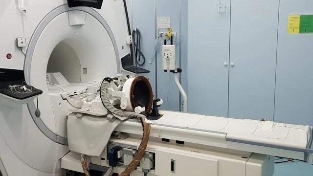
The technique used is focused ultrasound guided by magnetic resonance imaging (MRgFUS). This is a minimally invasive procedure with a device that triggers the ultrasounds affecting the brain (figure 2).
![Figure 2: MRgFUS machine [Source: University of L'Aquila].](/wp-content/uploads/sites/4/2020/04/macchinarioMRgFUS-eet.jpg)
The high-intensity waves are guided by an MRI and focused on a particular area of brain tissue. Once in the brain tissue, they heat and destroy the thalamus with a long-term reduction in tremors. The thalamus is an area of the brain below the cortex. Its role is fundamental and sophisticated: it selects and controls incoming information in relation to each experience.
The research team found that 37 out of 39 patients showed an immediate reduction in tremor. Even after some time, the tremor was lower. Some undesirable effects affected some patients, in particular limb weakness, and an alteration in the sensitivity of other parts of the body.
Currently, the only problems with this revolutionary procedure are that it is little-known and there are still few centers offering it, as Professor Bruno said: “The clinical application of this technique for neurological diseases is an absolute novelty; its clinical use was approved by the FDA less than three years ago. Few patients are aware of this therapeutic option, and there are still a few specialized centers that can offer it.”
The technique does not require invasive interventions. The brain tissue is heated in a targeted manner and then destroyed — through ultrasonic beams, safeguarding the surrounding healthy tissue. The technique presents less risk of bleeding or infection-related complications than deep brain stimulation or surgery to implant small electrodes. The disadvantage is that the process is irreversible, and any side effects are permanent.